Chest Wall Tumors
Chest Wall Tumors
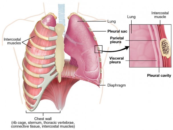
The chest cavity—which houses the lungs, heart, and other vital body parts—is a bone and muscle cage that is framed by the sternum (breastplate), spine, and ribs. The wall of the chest cavity provides rigid protection for your heart, lungs, and liver, but also is flexible to aid in respiration. It is made up of structures, including the rib cage, diaphragm, and abdomen. Like any other part of your body, your chest wall is susceptible to tumors.
Cells are the building blocks that make up the tissue and organs in your body. Old or damaged cells usually die and new cells take their place. But sometimes, the process goes wrong or a single cell starts to grow uncontrollably until it becomes a mass of tissue called a tumor.
Sometimes, tumors are non-cancerous (benign) and are not life threatening; other times, the tumors are cancerous (malignant) and can invade surrounding tissues or spread to other areas of the body.
Types Of Chest Wall Tumors
Tumors can arise from any different type of cell, including bone, muscle, and nerve cells. Non-cancerous chest wall tumors are relatively common and are treated only when they cause problems, such as breathing difficulties or pain. Cancerous chest wall tumors are rare and must be treated.
Non-cancerous tumors include:
- osteochondroma
- chondroma
- fibrous dysplasia
These types of tumors tend to run in families, but most present no cause for alarm and often remain undetected.
Cancerous tumors are usually sarcomas, which originate from soft tissue, cartilage, and/or bone in the chest. These cancerous tumors are categorized into two groups, depending on their origin:
- primary
- secondary
Primary tumors begin in the chest. Secondary tumors have spread (metastasize) from other parts of the body or directly extend into the chest wall from an adjacent breast or lung cancer.
Watch as Jules Lin, MD, explains more about chest wall tumors
Causes And Symptoms Of Chest Wall Tumors
- Chest Pain
- Swelling
It is unclear what causes chest wall tumors, although diet, lifestyle choices, and hereditary factors are believed to play a role.
Soft-tissue chest wall tumors don’t usually cause symptoms until the tumor is advanced. Tumors that are made up of cartilage or bone often cause pain, swelling, and impaired movement.
Chest x-rays can show whether or not you have an abnormality in your chest wall; however, x-rays are usually unable to distinguish benign tumors from malignant tumors.
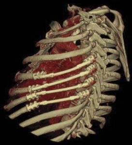
Diagnosis And Treatment Options
Computed tomography (CT) scan and magnetic resonance imaging (MRI) usually can zero in on the location and size of the tumor, as well as give some information about what type of tumor it is.
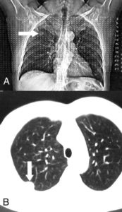
CT scan reveals tumor in the chest wall (arrows)
In the below video, Jules Lin, MD provides additional information on tests that can help diagnose a chest wall tumor and determine its size and location.
After a chest wall tumor is detected, your physician may want a biopsy to get more information about what type of tumor you have.
The most common procedure is an aspiration biopsy, where a needle is inserted into the tumor and cells are removed for examination. If it is too difficult to reach the tumor with a needle, you may need to undergo an open biopsy, which requires a small surgical incision and may leave a scar.
Your treatment plan will take into account the type of chest wall tumor you have, its size, and location. Malignant chest wall tumors may require a combination of chemotherapy, radiation therapy, and/or surgery.
If surgery is required, you also may need chest wall (ribs) or soft-tissue (muscle and skin) reconstruction so that your chest looks and functions normally.
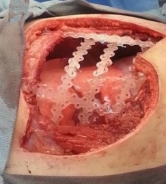
Examples of chest wall reconstruction.
As with any surgery or procedure, there are risks involved.
Recovery
Chest wall resections (removal of any chest wall structure) often cause more pain and require a longer healing period than other chest operations.
Talk with your doctor or cardiothoracic surgeon about the best treatment options for you.